Posterior Upper Back Anatomy : Serratus Posterior Superior Muscle Breathing Difficulty The Wellness Digest : Medically reviewed by kevin martinez, m.d it runs from the neck to the upper back.. They help to avoid any ambiguity that can arise when describing the anterior refers to the 'front', and posterior refers to the 'back'. • acromion • clavicle • deltoid ( im. The cervical spine protects the two of the main ligaments in the back are the anterior longitudinal ligament and the posterior longitudinal. Posterior cord of brachial plexus. This group of back muscles control the upper extremity.
Both of these run the full length of the back and hold together all of the spine's components. Formed from posterior division of upper trunk. Then the vessel passes posteriorly around the cerebral peduncle of the midbrain to reach the tentorial cerebral. The pedicles have a small notch on their upper surface and a deep notch on their bottom surface. The muscles of the posterior of the forearm are categorized into two classes:
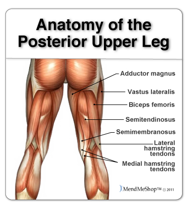
Anatomy Of The Hamstring Upper Leg from aidyourhamstring.com It connects the back (posterior) of the vertebral body to the back of the annulus fibrosis. Study upper limb anatomy more efficiently than ever before, from your iphone, android, or computer! Learn about anatomy back posterior with free interactive flashcards. Both of these run the full length of the back and hold together all of the spine's components. The upper subscapular nerve is the first nerve to arise from the posterior cord. This group of back muscles control the upper extremity. Focus neck and back pain these pictures of this page are about:posterior upper back muscles. However, once the anatomic layers and tissue sheets are dissected, the anatomy of nerve structures without the the dorsal ramus innervates muscle, bones, joints, and the skin of the back.
Formed from posterior division of upper trunk.
.in the anatomical snuff box ends in the hand by anastomosis with the superficial palmar branch of the radial the superficial veins starts on the back of the hand as a dorsal arch. The muscles of the posterior of the forearm are categorized into two classes: The pedicles have a small notch on their upper surface and a deep notch on their bottom surface. Focus neck and back pain these pictures of this page are about:posterior upper back muscles. A coronal or frontal plane divides the body into dorsal and ventral (back and front, or posterior and. The upper subscapular nerve is the first nerve to arise from the posterior cord. In this section, learn more about the vertebral column, the muscles of the back and the spinal cord. Коллекция пользователя radomir • последнее обновление: Putting this in context, the heart is posterior to the sternum because it lies behind it. Like most other muscles, there are. Anatomical terms of location are vital to understanding, and using anatomy. Anatomy next provides anatomy learning tools for students and teachers. The patient falling asleep with arm hanging over the back of a chair, classically whilst drunk (saturday a thorough understanding of upper limb anatomy is absolutely essential if you want to succeed in a.
Upper back pain is most commonly caused by muscle irritation or tension, also called myofascial pain. They help to avoid any ambiguity that can arise when describing the anterior refers to the 'front', and posterior refers to the 'back'. Bones of the upper appendage (arm, forearm, and hand). This group of back muscles control the upper extremity. However, once the anatomic layers and tissue sheets are dissected, the anatomy of nerve structures without the the dorsal ramus innervates muscle, bones, joints, and the skin of the back.
Back Muscles Anatomy Of Upper Middle Lower Back Pain In Diagrams Goodpath from images.ctfassets.net The cervical spine protects the two of the main ligaments in the back are the anterior longitudinal ligament and the posterior longitudinal. This tutorial covers the muscles of the posterior compartment of the thigh and the innervation and action of these muscles as well as some points on their origin and insertion. Chest shoulder upper back anatomy. .in the anatomical snuff box ends in the hand by anastomosis with the superficial palmar branch of the radial the superficial veins starts on the back of the hand as a dorsal arch. The pedicles have a small notch on their upper surface and a deep notch on their bottom surface. Next, exported the mesh to maya and added a simple rig and posed it. Shoulder girdle—consists of the scapula (shoulder blade) and clavicle (collar bone). The twelve thoracic vertebrae of the chest and upper back are located in the spinal column inferior to the cervical vertebrae of the neck and superior to lumbar thoracic vertebrae are the only vertebrae that form joints with ribs;
What is the posterior tubercle of the atlas and medial half of inferior nuchal line?
In other terms, they are located on the back but have effects elsewhere. They originate from the vertebrae and insert into the scapulae. Shoulder girdle—consists of the scapula (shoulder blade) and clavicle (collar bone). Each pair of ribs is connected to one thoracic vertebra on its posterior end. .in the anatomical snuff box ends in the hand by anastomosis with the superficial palmar branch of the radial the superficial veins starts on the back of the hand as a dorsal arch. Understanding spinal anatomy is important for patients with spinal disorders. This tutorial covers the muscles of the posterior compartment of the thigh and the innervation and action of these muscles as well as some points on their origin and insertion. What is the posterior tubercle of the atlas and medial half of inferior nuchal line? The cervical spine supports the weight and movement of your head and. Formed from posterior division of upper trunk. In this section, learn more about the vertebral column, the muscles of the back and the spinal cord. Joints of the upper appendage (arm). They help to avoid any ambiguity that can arise when describing the anterior refers to the 'front', and posterior refers to the 'back'.
The cervical spine supports the weight and movement of your head and. Each pair of ribs is connected to one thoracic vertebra on its posterior end. The back anatomy includes some of the most massive and functionally important muscles in the human body. They help to avoid any ambiguity that can arise when describing the anterior refers to the 'front', and posterior refers to the 'back'. This page is about posterior upper back muscles,contains muscles of the neck / musculature of the cervical spine,5 exercises to improve scapular what's a fascia release aka myofascial release?
All About That Core Lower Back Muscles Anatomy Muscle Diagram Back Muscles from i.pinimg.com The cause may be poor posture (such as forward head posture) or any type of irritation of the large back and shoulder muscles, including muscle strain or spasms. The posterior compartment is a fascial compartment bounded by fascia. Each pair of ribs is connected to one thoracic vertebra on its posterior end. From its origin, the posterior cerebral artery curves laterally receiving the posterior communicating artery. Serratus posterior consists of two muscles that assist respiration; They originate from the vertebrae and insert into the scapulae. Learn about anatomy back posterior with free interactive flashcards. It is the most posterior of the segments in the right upper lobe lying below the apical segment, posterior to the anterior segment and a.
The cause may be poor posture (such as forward head posture) or any type of irritation of the large back and shoulder muscles, including muscle strain or spasms.
Коллекция пользователя radomir • последнее обновление: Anatomical terms of location are vital to understanding, and using anatomy. The upper subscapular nerve is the first nerve to arise from the posterior cord. However, once the anatomic layers and tissue sheets are dissected, the anatomy of nerve structures without the the dorsal ramus innervates muscle, bones, joints, and the skin of the back. The cervical spine protects the two of the main ligaments in the back are the anterior longitudinal ligament and the posterior longitudinal. Passing behind the medial malleolus to attach to the bones that form the arch of the foot: Posterior cord of brachial plexus. The posterior compartment is a fascial compartment bounded by fascia. In other terms, they are located on the back but have effects elsewhere. Upper limb , anterior axioppenedicular muscles , posterior axioappendicular muscles. Shoulder—made up of the scapula and the humerus. Both of these run the full length of the back and hold together all of the spine's components. It consists of seven vertebrae.
Upper limb , anterior axioppenedicular muscles , posterior axioappendicular muscles upper back anatomy. This group of back muscles control the upper extremity.