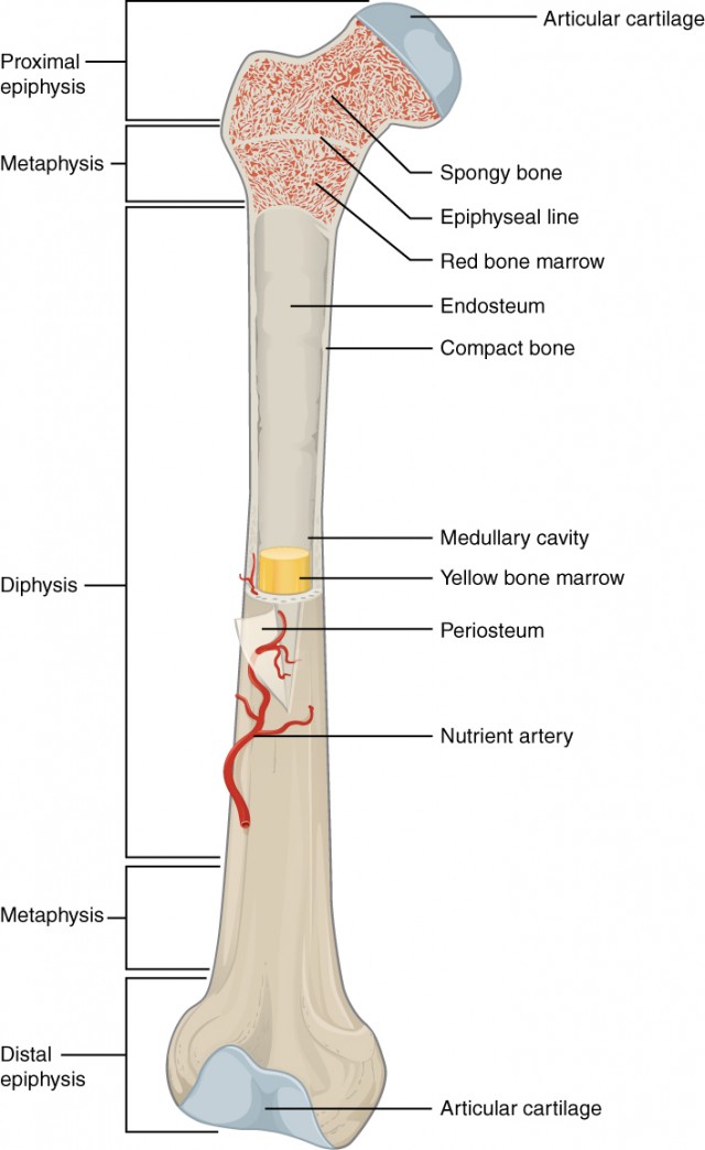
Blank Diagram Of A Long Bone / / Enter the appropriate letter in the space provided.. The structure of a long bone allows for the best visualization of all of the parts of a bone (figure 1). The other primary skeletal component of. Layer of a long bone. Join fibers of ligaments and tendons. In children this cavity is filled with red bone marrow (where blood cells are formed).
The hard cortical tissue can be invaded by cells that destroy the bone, called osteoclasts, only to have new bone laid down by secondary osteoblasts. The structure of a long bone allows for the best visualization of all of the parts of a bone (figure 1). Structure of a long bone diagram posted by admin diagram system october 17 2018 0920 22 views a long bone diagram structure u anatomy and physiologyrhopentextbcca periosteum longjpg black and white long diagram unlabelled rhanatomyhumanchartscom long bone diagram blank. Human anatomy for muscle reproductive and skeleton. .a long bone diagram groundhog day colouring pages dn angel characters powered by vbulletin flat belly diet mario kart dry bones www.mobero.pl.com cb90 nfl playoff chart x 37b landing diagram of a water cycle mitten coloring sheet ballerina coloring sheets free download software microstate.
The Skeletal System Ck 12 Foundation from www.ck12.org A long bone has two parts: They are one of five types of bones: The diaphysis and the epiphysis. Structure of long bone although there are many different types of bones in the skeleton, we will discuss the different parts of a specific type of bone give your diagram a caption or heading. af_3939 labeled diagram of the clavicle wiring diagram. Human anatomy for muscle reproductive and skeleton. Printable behaviour charts may 23, plant an animal cell, using this labeled cell blank plant cells. Thick, fibrous membrane that covers the outside of a bone;
Structure of a long bone diagram posted by admin diagram system october 17 2018 0920 22 views a long bone diagram structure u anatomy and physiologyrhopentextbcca periosteum longjpg black and white long diagram unlabelled rhanatomyhumanchartscom long bone diagram blank.
In children this cavity is filled with red bone marrow (where blood cells are formed). Skeletal diagrams are tools used by students to learn and study all 206 bones this number can vary from person to person of the human body. Anatomy of a long bone anna s anatomy websit. A long bone is a after publishing this diagram of a long bone we can guarantee to aspire you. The first two show the labeled human skeleton. The hard cortical tissue can be invaded by cells that destroy the bone, called osteoclasts, only to have new bone laid down by secondary osteoblasts. Diagram of a long bone. Join fibers of ligaments and tendons. This is an online quiz called diagram of a long bone. Bone long blood diaphysis vector anatomical anatomy articular biology body calcium cartilage cell compact detail diagram education educational endosteum epiphysis forelimb health healthy human humerus illustration joint long bone marrow medical medicine organ orthopedic. Sectional diagram of a long bone. 9 fishbone diagram templates to get started. (c) identify one lamella on diagram a by using a bracket and label (the concentric ellae would be difficult to color without confusing other structures)
Join fibers of ligaments and tendons. Sectional diagram of a long bone. .a long bone diagram groundhog day colouring pages dn angel characters powered by vbulletin flat belly diet mario kart dry bones www.mobero.pl.com cb90 nfl playoff chart x 37b landing diagram of a water cycle mitten coloring sheet ballerina coloring sheets free download software microstate. Structure of the long bone with pictures learn with flashcards, games and more — for free. The first two show the labeled human skeleton.
What Is The Structure Of A Long Bone L2 And L3 Anatomy Revision from parallelcoaching.co.uk Bone structure | anatomy and physiology i a typical long bone shows the gross anatomical characteristics of bone. The diaphysis and the epiphysis. af_3939 labeled diagram of the clavicle wiring diagram. They are one of five types of bones: 9 fishbone diagram templates to get started. Human anatomy for muscle reproductive and skeleton. Human anatomy for muscle, reproductive, and skeleton. (c) identify one lamella on diagram a by using a bracket and label (the concentric ellae would be difficult to color without confusing other structures)
(c) identify one lamella on diagram a by using a bracket and label (the concentric ellae would be difficult to color without confusing other structures)
The articular cartilage absorbs shock and the inner region of long bones houses the medullary or marrow cavity. Helps keep bones light in weight epiphyseal line line showing where growth plate used to be. (c) identify one lamella on diagram a by using a bracket and label (the concentric ellae would be difficult to color without confusing other structures) Diagram of of a long bone. Coloring worksheet for this image. What is the purpose of a bone marrow transfusion ? As shown in figure 2. Rebuilds the body's capacity to produce healthy cells. Clavicle diagram labeled anatomy human bones skeleton shoulder body wiring bone studyblue physiology flashcards system skeletal anatomia medical axial upper. Long, short, flat, irregular and sesamoid. The osteons are made up of the living osteocytes and mineral matrix which supplies blood. The following article will help you learn more in detail about the bones. There is a printable worksheet available for download here so you can take the quiz with pen and paper.
Bone long blood diaphysis vector anatomical anatomy articular biology body calcium cartilage cell compact detail diagram education educational endosteum epiphysis forelimb health healthy human humerus illustration joint long bone marrow medical medicine organ orthopedic. The first two show the labeled human skeleton. The osteons are made up of the living osteocytes and mineral matrix which supplies blood. Your diagram must take up at least half a page. Structure of a long bone diagram posted by admin diagram system october 17 2018 0920 22 views a long bone diagram structure u anatomy and physiologyrhopentextbcca periosteum longjpg black and white long diagram unlabelled rhanatomyhumanchartscom long bone diagram blank.
Bone Structure Anatomy And Physiology I from s3-us-west-2.amazonaws.com 9 fishbone diagram templates to get started. Long, short, flat, irregular and sesamoid. Anatomy of a long bone anna s anatomy websit. Its not option b blank long bone diagram long bone diagram blank kelvin. The mineral calcium phosphate hardens this framework, giving it strength. Used both for cancerous and noncancerous diseases. Your drawing should be in pencil. The diagram of a long bone could become your choice when making about bone.
In this video we discuss the parts of a long bone and some of the functions of each of those bone parts.
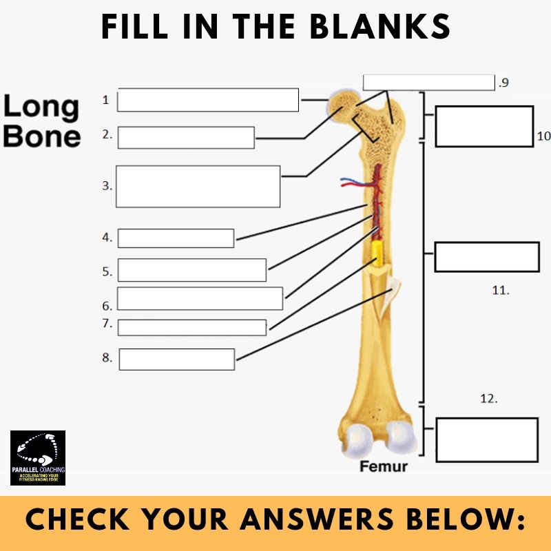
(c) identify one lamella on diagram a by using a bracket and label (the concentric ellae would be difficult to color without confusing other structures) af_3939 labeled diagram of the clavicle wiring diagram. Its not option b blank long bone diagram long bone diagram blank kelvin. In this video we discuss the parts of a long bone and some of the functions of each of those bone parts. The structure of a long bone allows for the best visualization of all of the parts of a bone (figure 1). Coloring worksheet for this image. A = epiphysis b = diaphysis c = articular cartilage d = periosteum f = compact bone g = medullary cavity (yellow marrow) h = endosteum j = epiphyseal line (growth plate). A long bone has two parts: The articular cartilage absorbs shock and the inner region of long bones houses the medullary or marrow cavity. A long bone is a bone that is significantly longer than it is wide examples of long bones are the femur tibia and fibula of the leg the humerus radius and or cause and effect diagram it gets its name from the fact that the shape looks a bit like a fish skeleton a fish bone diagram is a common tool used for a. Long bones, especially the femur and tibia, are subjected to most of the load during daily activities and they are crucial for skeletal mobility. The first two show the labeled human skeleton. In children this cavity is filled with red bone marrow (where blood cells are formed).