Upper Leg Tendon Anatomy - - It is formed when the soleus muscle tendon joins with the gastrocnemius tendon.. Collectively, they act to dorsiflex and invert the foot at the ankle joint. The thigh bone, or femur, is the large upper leg bone that connects the lower leg bones (knee joint) to the pelvic bone (hip joint). Plantar flexion of the foot, ankle joint stabilizer. Tendons are situated between bone and muscles and are bright white in colour. Leg muscle anatomy chart | amulette.
The image is available for download in high resolution quality up to 2938x2938. The thigh bone, or femur, is the large upper leg bone that connects the lower leg bones (knee joint) to the pelvic bone (hip joint). This mri wrist coronal cross sectional anatomy tool is absolutely free to use. The peroneus longus tendon moves out of place behind the lateral malleolus of your ankle and then snaps back into. Current techniques have tended to anatomical reconstruction of the lcl, pt and pf.
Thigh Pain Causes Treatment And When To See A Doctor from www.verywellhealth.com Marc draws and describes the form and location of the upper leg front position. .16 penile numbness and perineum tenderness.18 any suggested exercises or stretches?.22 leg musculature 209 elbow tendonitis and saddle sores. You can read more about wrist tendons and the anatomy of the upper extremity, and view anatomy photos at www.handcare.org. Lie prone on a hamstring curl machine. 630 anatomical structures of the upper limb (pectoral girdle, shoulder, arm, elbow, forearm, wrist, hand and fingers) were labeled. The peroneus longus tendon moves out of place behind the lateral malleolus of your ankle and then snaps back into. ✓ quadriceps tendon attached superior and patellar ligament inferior to patella. Hands are outstretched, holding onto the handles of the bench.
Superficial veins of upper limb , anatomy :
It attaches the calf muscles to the calcaneus (heelbone) and allows us most of the motion of the ankle is caused by the stronger muscles in the lower leg whose tendons pass by the ankle and connect in the foot. 1280 x 1520 jpeg 166 кб. Use the mouse scroll wheel to move the images up and down alternatively use the tiny arrows (>>) on both side of the image to move the images. Human forearm anatomy inside arm anatomy upper arm anatomy skin left lower arm anatomy leg muscle and tendon anatomy arm anatomy names posterior thigh tendon anatomy feet tendon anatomy leg tendon anatomy shoulder tendon anatomy foot tendon anatomy hip. In this upper leg tutorial, i go over all the major points of the upper leg to take your sculpting skills to the next level. Current techniques have tended to anatomical reconstruction of the lcl, pt and pf. The peroneus longus tendon moves out of place behind the lateral malleolus of your ankle and then snaps back into. Leg anatomy muscles and tendons how to fix achilles. This article will discuss the anatomy and function of the achilles tendon. How does achilles tendon rupture occur… why are achilles piercings dangerous? It is formed when the soleus muscle tendon joins with the gastrocnemius tendon. Related posts of muscle anatomy upper leg. Horse leg basic anatomy tendons подробнее.
Lateral (fibular) collateral ligament (fcl) upper part middle part lower part popliteus tendon (pt) upper part i. There are four muscles in the anterior compartment of the leg. Horse leg basic anatomy tendons подробнее. Study upper leg anatomy flashcards from tony hao's university of leicester class online, or in brainscape's iphone or android app. The lower leg is comprised of two bones, the tibia and the smaller fibula.
Muscular Function And Anatomy Of The Upper Leg Video Lesson Transcript Study Com from study.com They are remarkably strong, having one of the highest tensile strengths found among soft tissues. The thigh bone, or femur, is the large upper leg bone that connects the lower leg bones (knee joint) to the pelvic bone (hip joint). ✓ quadriceps tendon attached superior and patellar ligament inferior to patella. Muscle/tendon inflammation and pain along anterio… Lateral (fibular) collateral ligament (fcl) upper part middle part lower part popliteus tendon (pt) upper part i. Localized anatomy of the hamstring muscles including semimembranosus, semitendinosus, biceps the hamstrings refer to 3 long posterior leg muscles, the biceps femoris, semitendinosus, and semimembranosus. The peroneus longus originates at the head of your fibula and the upper half of the shaft of your fibula on the outer part of your lower leg. There is no real division between the core and the upper leg;
Tendons transmit the mechanical force of muscle contraction to the bones.
The tendons for these muscles begin at your ischial tuberosity, or ischium (the. Hands are outstretched, holding onto the handles of the bench. Muscle/tendon inflammation and pain along anterio… The large achilles tendon is the most important tendon for walking, running, and jumping. Use the mouse scroll wheel to move the images up and down alternatively use the tiny arrows (>>) on both side of the image to move the images. Leg muscle anatomy chart | amulette. Common tendon of superficial posterior leg muscles; This may result in tendon subluxation; Lie prone on a hamstring curl machine. Horse leg basic anatomy tendons подробнее. Tendon, tissue that attaches a muscle to other body parts, usually bones. The tendons that control movement in your hands, wrists and fingers run through your forearm. The tendons of the edl can be palpated on the dorsal surface of the foot.
Hands are outstretched, holding onto the handles of the bench. Muscle/tendon inflammation and pain along anterio… It is formed when the soleus muscle tendon joins with the gastrocnemius tendon. Leg muscle anatomy chart | amulette. Leg anatomy muscles and tendons how to fix achilles.
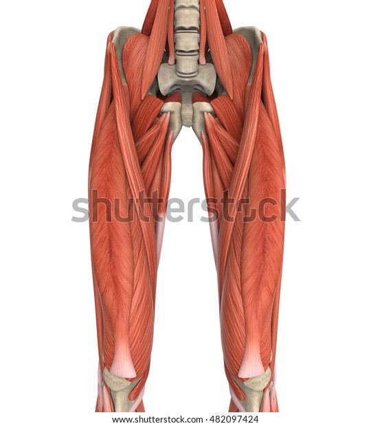
Upper Legs Muscles Anatomy 3d Rendering Stock Illustration 482097424 from image.shutterstock.com The lower leg is comprised of two bones, the tibia and the smaller fibula. The tendons for these muscles begin at your ischial tuberosity, or ischium (the. Lateral (fibular) collateral ligament (fcl) upper part middle part lower part popliteus tendon (pt) upper part i. Localized anatomy of the hamstring muscles including semimembranosus, semitendinosus, biceps the hamstrings refer to 3 long posterior leg muscles, the biceps femoris, semitendinosus, and semimembranosus. This mri wrist coronal cross sectional anatomy tool is absolutely free to use. Originates from the lateral condyle of the tibia and the medial surface of the fibula. Palmar region , arteries (illustrations: The calcaneal tendon, also known as the tendon of achilles, is a posterior leg tendon — a fibrous connective tissue that joins muscles in the back of the leg.
The human leg, in the general word sense, is the entire lower limb of the human body, including the foot, thigh and even the hip or gluteal region.
The human leg, in the general word sense, is the entire lower limb of the human body, including the foot, thigh and even the hip or gluteal region. Hands are outstretched, holding onto the handles of the bench. Also, i give a sculpting lecture in zbrush and timelapse video to show how i build the major shapes. Plantar flexion of the foot, ankle joint stabilizer. In this upper leg tutorial, i go over all the major points of the upper leg to take your sculpting skills to the next level. The tendons of the edl can be palpated on the dorsal surface of the foot. There is no real division between the core and the upper leg; Superficial veins of upper limb , anatomy : Current techniques have tended to anatomical reconstruction of the lcl, pt and pf. Common tendon of superficial posterior leg muscles; 1280 x 1520 jpeg 166 кб. ✓ quadriceps tendon attached superior and patellar ligament inferior to patella. This may result in tendon subluxation;
/thighpainfinal-01-5c1b07c8c9e77c0001fed2d4.png)